Diabetic foot disease often develops as a “chronic accumulation,” with early symptoms being subtle, but the plantar structure, pressure, temperature, and blood flow status have already quietly changed. Traditional health examination methods mainly rely on visual observation or physician palpation, which makes it difficult to detect these small differences. However, the foot scanner based on laser scanning three-dimensional data brings a new quantitative approach to diabetic foot risk assessment.
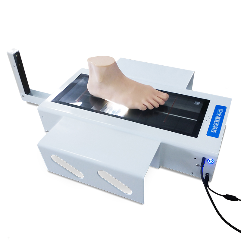
The laser foot scanner uses a high-precision laser ranging system to perform comprehensive non-contact scanning of the plantar surface. During the scanning process, the laser beam rapidly moves to the plantar surface, measuring the spatial coordinates of each point through reflection time or angle differences, and then creating a complete three-dimensional foot model. This process can be completed in a few seconds, with a resolution that can reach submillimeter levels, accurately reconstructing the complex structures of the arch, toes, and heel.
The three-dimensional foot model is not just a shape diagram; it is also a “health record.” By comparing changes in height, curvature, and pressure characteristics at different parts of the foot, the system can automatically identify abnormal points. For example, due to nerve damage in diabetic patients, some areas may have reduced sensation but bear more pressure, which over time can easily lead to pressure ulcers. The three-dimensional data can precisely reflect the locations and areas of these high-pressure zones, providing quantitative evidence for doctors.
Additionally, when combined with pressure analysis modules, laser scanning can overlay dynamic pressure data of the foot, forming a “morphology and mechanics” dual-dimensional evaluation. Doctors can clearly identify which areas are subjected to excessive pressure and which areas lack necessary support. Long-term comparison of scanning results at different stages can also detect early signs of foot arch collapse and toe bone deformation, enabling intervention before ulcers occur.
Laser scanning technology also has significant advantages such as repeatability and objectivity. The results measured at different times by different doctors can be highly consistent. This is especially important in chronic risk monitoring, such as for diabetic foot. Patients can undergo regular scans, and by analyzing the trends of three-dimensional data, doctors can determine the progression of the disease, such as the descent of the arch, the rise of metatarsal bones, or the atrophy of subcutaneous tissue, all of which can be automatically identified and risk-flagged by the system.
At the same time, the scanning data can be linked to the hospital information system to build a personal foot health database. Doctors can observe the static morphology and also integrate blood sugar, blood flow, and temperature for multidimensional analysis, even using artificial intelligence algorithms to predict future high-risk areas. This transforms the foot scanner from a “measuring tool” into a “risk monitoring platform,” achieving the goals of early detection and early intervention.
